What is a PET scan?
A positron emission tomography (PET) scan is an imaging test that generates images of the functioning of your organs and tissues. The test utilizes an injectable radioactive chemical known as a radiotracer and a PET scanner.
The scanner detects diseased cells that absorb substantial quantities of the radiotracer, indicating the presence of a potential health issue.
PET scans are frequently utilized to aid in cancer diagnosis and treatment evaluation. Using the scan, they can also evaluate particular heart and cerebral conditions.
What is the difference between PET, CT, and MRI scans?
X-rays are used in computed tomography (CT) examinations. Magnets and radio waves are utilized for magnetic resonance imaging (MRI) tests. Both produce images of organs and skeletal structures.
PET scans use a radioactive tracer to demonstrate the real-time function of an organ. PET scan images can detect cellular changes in organs and tissues before CT and MRI scans. Your doctor may perform a PET and CT scan simultaneously (PET-CT). This combination test generates 3D images for a more precise diagnosis.
Some institutions now utilize PET/MRI hybrid scans. This novel technology produces images with extremely high contrast. This scan is primarily used for diagnosing and monitoring soft tissue cancers (brain, head and neck, liver, and pelvis).
What is a PET scan used to detect?
Your doctor may prescribe a PET scan to check for the presence of:
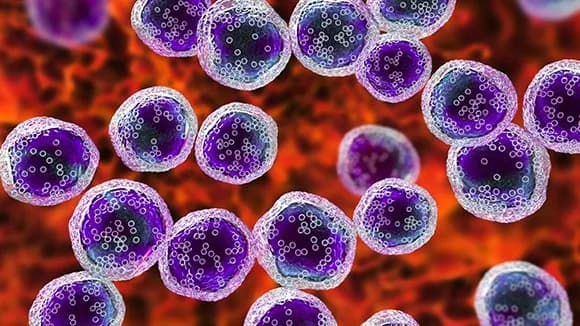
- Cancer, including breast, lung, and thyroid cancer.
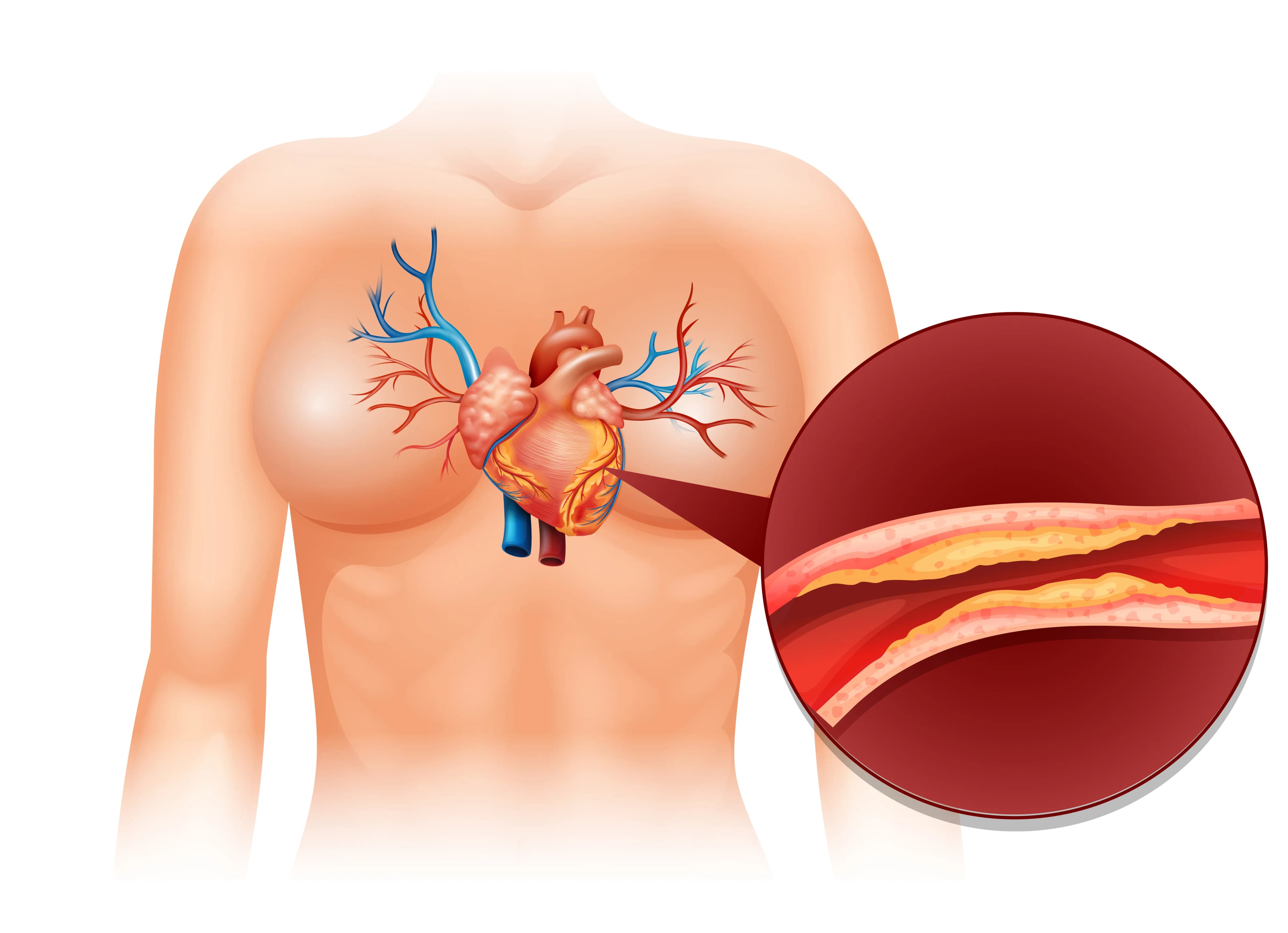
- Coronary artery disease, heart attack, or other cardiovascular issues
- Brain disorders such as brain tumours, epilepsy, dementia, and Alzheimer's disease.
How the Test is Performed
PET scans utilize a radioactive tracer. The tracer is administered intravenously (IV). Typically, the needle is inserted in the inner forearm. The tracer circulates in the circulation and accumulates in organs and tissues. This enables the radiologist to see specific areas with greater clarity.
You must wait for the tracer to be assimilated by your body. This takes roughly one hour.
You will then lie on a narrow table gliding into a large tunnel-shaped scanner. The PET detects the tracer's signals. A computer converts signals into three-dimensional images. The images are displayed on a monitor for your physician to view.
You must remain still throughout the exam. Movement can obscure images and result in errors.
The duration of the examination depends on which body region is being scanned.
How to Prepare for the Test
You may be instructed to fast for four to six hours before the scan. You may only consume water, not other beverages, including coffee. If you have diabetes, your doctor will instruct you not to take your diabetes medication before the exam. These medications will impede the results.
Inform your provider if:
- You fear confined spaces (suffer from claustrophobia). You may be prescribed a medication to induce sleep and reduce anxiety.
- You are pregnant, or you suspect that you may be pregnant.
- You have an allergy to the contrast dye that will be injected.
- Always inform your healthcare provider of the medications you are consuming. Inform your provider of any medicines you purchased without a prescription. Occasionally, medications may interfere with test results.
How the Test will Feel
- When the tracer catheter is inserted into your vein, you may experience a sharp pinch.
- A PET scan is painless. If the table is chilly or uncomfortable, you can request a blanket or pillow.
- The room's intercom enables you to communicate with anyone at any time.
- There is no recovery period unless a sedative is administered.