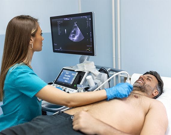
What is 2-D Echocardiography?
2-D echocardiography, commonly referred to as 2-D echo, is a non-invasive diagnostic procedure employed for evaluating the performance and examining the various segments of the heart. Utilizing sound vibrations, this test generates images of distinct heart components, aiding in examining damages, obstructions, and blood flow rates. Regular 2-D echo examinations are advised by physicians to detect and address potential heart issues in their early stages, contributing to maintaining good health and vitality as you age.
Why might I need an Echocardiography?
2-D echocardiography is conducted to identify the following cardiac conditions:
1. Any hidden heart diseases or irregularities.
2. Congenital heart diseases, blood clots, or tumours
3. Dysfunction of the heart valve.
4. Abnormal blood flow within the heart.
The information obtained through 2-D echo pertains to the heart's functionality, aiding in diagnosing malfunctions and formulating treatment plans for emerging diseases.
Risks of 2-D echocardiography
This imaging technique is non-invasive and involves minimal to no risks. Discomfort may arise due to the transducer's positioning, exerting pressure on the body's surface. Some individuals may experience discomfort or pain from lying still on the examination table throughout the procedure. Additional risks may be present based on individual health conditions, and addressing any concerns with your doctor before undergoing the procedure is advisable.
How to get ready for 2-D echocardiography?
1. Your physician will provide an overview of the procedure and ask if you have any questions.
2. Typically, preparation, such as fasting or sedation, is not required.
3. Inform your doctor about all prescribed and over-the-counter medications and herbal supplements you are currently using.
4. Let your doctor know if you have a pacemaker.
5. Depending on your medical condition, your doctor may recommend additional specific preparations.
What happens during the procedure?
The procedure lasts approximately 30 minutes to an hour and is conducted safely under the supervision of a radiologist and a cardiologist.
1. During the process, the lab technician applies a colourless gel, and the transducer is moved across different areas of the chest to assess sound vibrations.
2. The gel aids the transducer in obtaining views of the heart, tissues, and structures.
3. Following the test, the gel is wiped off.
You will be provided with test images either in print or recorded on a DVD, allowing your physician to interpret the results for any abnormalities or potential developing diseases.
What happens after the procedure?
Unless otherwise instructed by your doctor, please return to your regular diet and activities.
Typically, there is no specific post-echocardiogram care required. However, your doctor may provide additional instructions based on your circumstances following the procedure.
Secure the well-being of your heart and contribute to your family's happiness by scheduling our basic or advanced cardiac package today. Take advantage of our discounted prices and book your preferred package without delay.
Speak to our team about 2-D echocardiography
We encourage you to speak with our knowledgeable team if you have questions or concerns about 2-D echocardiography. Our team can provide information about the procedure, its benefits, and how it can contribute to understanding your heart health. Whether seeking clarification or considering undergoing a 2-D echocardiogram, our experts address your inquiries and ensure you have the information you need for informed decision-making. Feel free to contact us to discuss 2-D echocardiography and its role in assessing and monitoring your cardiac well-being.
FAQS
Health In A Snap, Just One App.
KNOW MORE
