
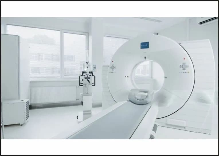
Medical technology has made remarkable strides in recent years, transforming the way doctors diagnose and treat diseases. One such breakthrough is the PET-CT scan, which has become an essential tool in modern healthcare. In fact, studies suggest that more than 2 million PET scans are performed globally every year, helping in the early detection and monitoring of conditions ranging from cancer to neurological disorders.
With its ability to deliver detailed insights into organ function and disease progression, a PET-CT scan empowers doctors to create more precise and personalized treatment plans. In the following sections, we’ll explore what a PET-CT scan is, how it works, how it compares to other imaging methods, and why it has become such a vital tool in healthcare today.
A Positron Emission Tomography (PET) scan is a powerful imaging test that provides detailed pictures of how your organs and tissues are working, not just how they look. Unlike standard scans that show structure, PET scans reveal the metabolic and biochemical activity inside your body.
The test involves a safe, injectable radioactive substance known as a radiotracer (or tracer). Once injected, the tracer travels through your bloodstream and collects in areas of the body with higher levels of activity. Since diseased or cancerous cells often use more energy than normal cells, they absorb larger amounts of the tracer. This highlights potential health problems, sometimes before they appear on conventional imaging like CT (Computed Tomography) or MRI (Magnetic Resonance Imaging).
Healthcare providers commonly use PET scans to:
In many cases, PET images are combined with CT or MRI scans to provide even more precise information. A PET-CT scan merges the metabolic detail of PET with the structural clarity of CT, creating a comprehensive 3D image. This combination is especially valuable in cancer care, as it shows both the exact location and the activity level of abnormal cells.
During the procedure, the tracer is typically injected into a vein in your arm or hand. After a short waiting period, you’ll lie on a scanning table while the machine captures images. The scanning itself usually takes 30–45 minutes, though the entire process, from preparation to completion, can take around 2 hours. PET-CT scans are usually done in the radiology department as an outpatient procedure.
Because these scanners are highly specialized, they are often found in major cancer hospitals or advanced diagnostic centres, which may require some patients to travel to access the service. With its ability to detect disease activity at the cellular level, a PET-CT scan has become an indispensable tool in early diagnosis, treatment planning, and monitoring outcomes in modern medicine.
Doctors usually recommend a PET-CT scan for conditions that need both structural and functional insight.
Medical imaging plays a crucial role in diagnosing and monitoring diseases. While CT scans, MRI scans, and PET scans are all advanced imaging tools, each works differently and serves unique purposes in healthcare.
CT scan (Computed Tomography)
MRI (Magnetic Resonance Imaging)
PET Scan (Positron Emission Tomography)
Yes. The amount of radiation is carefully controlled and generally safe, even for repeated use when medically necessary. Your doctor will weigh the benefits against any potential risks before recommending it.
You should consider consulting your doctor for a PET-CT scan if you:
At Apex Hospitals, our advanced PET-CT technology helps doctors detect diseases early, monitor treatment accurately, and guide precise interventions. With expert radiologists, compassionate staff, and NABH-accredited facilities, we ensure world-class care.


Download Our App


Powered By Docfyn